ACIST HDi High-Definition IVUS Systemは、高度なイメージングモード、児玉IVUSカテーテルの配信可能性の向上、インタラクティブなコンパクトコンソールを通じて、冠状動脈および末梢の介入戦略に情報を提供し、照らすための最適化された視覚化を可能にします。
ACIST HDiの詳細については、担当者にお問い合わせください。
現在、冠状動脈および末梢血管の両方の手技に適応されています。
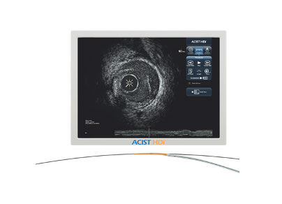
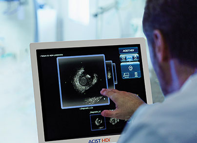
HDiと 強化されたイメージングモード 処置前の計画と処置後の評価のために、より明確に定義されたIVUSイメージを提供します1製品の利点インタラクティブ タッチスクリーン付きコンパクトコンソール
迅速な分析 簡単なキャスラボ統合のための小さな設置面積製品の利点納品性の向上2 最適化されたイメージング3 とと
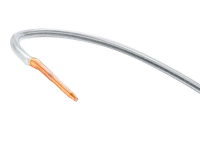
設置面積製品の利点納品性の向上2 最適化されたイメージング3 とともに遠位端をオフセット児玉のユニークな可変柔軟性(VariFlex™)イメージングウィンドウ® HDIVUSカテーテル
確実に見る力を提供します。
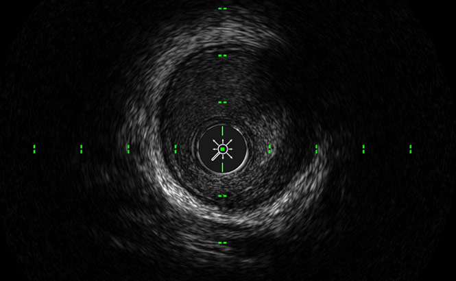
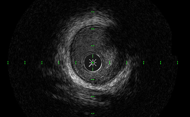
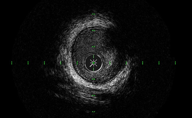
より良い境界検出のために冠状動脈内腔を暗くします。
グレースケールを大きくして、血液のスペックル、組織、プラークをより細かく区別します。
高解像度と浸透の深さのバランスを最適化し、完全な血管壁の視覚化を可能にします
臨床的証拠
より良い視覚化 ステントのサイジングを最適化するためのOCTよりもメディアの4プラーク量が多い場合でも、60 MHzで十分な浸透を提供してメディア層を確認することにより、イメージングを強化できます4、したがって、医師はステント留置断面積を最大化でき、より良い患者転帰につながる可能性があります5
IVUSの使用により、手技中に治療戦略が変更されました 74%の時間6IVUSガイド下ステントも近位および遠位のプラーク負荷が低く、MSAが高かった血管造影グループと比較して7
8倍以上 40MHzより60MHzでの脂質プールの検出8IVUSガイド下PCIは、血管造影のみと比較して、びまん性プラーク病変がある場合のステントエッジ再狭窄の予防に有益です9
IVUSは、より安価で費用効果が高い血管造影 PCI手順の71%10IVUSは、特にリスクの高いサブグループ(糖尿病、腎不全、ACS)でより大きな経済的利益をもたらし、より低いコストで改善された結果を提供します。これは、イタリアの医療費支払者の観点から見た生涯経済モデルに基づいています10
下肢処置におけるIVUSの使用は関連していた 合併症のリスクが低い 術後および切断のリスクの低下11
2.8倍少ない標的病変血管新生 血管造影のみと比較して、方向性アテローム切除術でIVUSを使用した場合の1年間のフォローアップ12
血管造影画像から得られた視覚的推定測定値は一貫して すべての動脈セグメントにわたって小さい IVUS評価から得られたものより13
カルシウムが69%多く検出されました同じ病変の血管造影と比較したIVUSによる14
2倍以上の血栓が特定された 血管造影と比較したIVUSによる15
ステント後の解剖の4倍 血管造影と比較してIVUSによって識別された163.5倍以上の血管形成術後の解剖 血管造影と比較してIVUSによって識別された17アテローム切除後の解剖の6倍 血管造影と比較してIVUSによって識別された17
ファイル上のデータ– TR-07057 –内部テストファイル上のデータ– TR-4050 –児玉カテーテル性能の研究概要ファイル上のデータ– TR-07057 –内部テストIVUSガイド下対OCTガイド下冠状動脈ステント留置: 重要な評価 https://doi.org/10.1016/j.jcmg.2017.09.008IVUS最適化薬剤溶出ステント留置の新しい基準の定義: PRAVIO研究。 カテーテルCardiovascInterv。 2009年8月1日; 74(2):348-356。 6.6。 前原A他– 2018年11月16日https://doi.org/10.1161/CIRCINTERVENTIONS.117.006243Circulation: 心臓血管の介入。 2018; 11:e006243https://www.tctmd.com/news/ultimate-ivus-superior-angiography-guiding-pci-less-tvf-1-year. https://doi.org/10.1016/j.jacc.2018.09.01 8.8。 田中聡、坂本晃、北原秀樹ほか 新しい高解像度60MHz IVUSイメージングシステムによる脂質プラークと血栓の評価: 従来の40MHzIVUSおよびOCTとの比較。 J Am Coll Cardiol 2013; 62(18_S1):B201-B2028a。 合計50の一致する断面が分析され、ex-vivo研究では8つの断面で脂質コアが特定されました。DES移植後のエッジ再狭窄に対するステントエッジから残存プラークまでの距離の影響。 PLOSOne。 2015; 10(3):E0121079。 10.10。 Alberti、A.、Giudice、P.、Gelera、A。etal。 血管内超音波(IVUS)の経済的影響を理解する。 Eur J Health Econ 17、185–193(2016)。 https://doi.org/10.1007/s10198-015-0670-4Panaich、S。 S.、アロラ、S。、パテル、N。、パテル、N。 J.、Savani、C.、Patel、A。、…Badheka、A。 O.(2016)。 下肢末梢血管インターベンションにおける血管内超音波: 全国の入院患者サンプル(2006年から2011年)からの利用率の変動と院内転帰への影響。 Journal of Endovascular Therapy、23(1)、65–75。 https://doi.org/10.1177/1526602815620780Krishnan、P.、Tarricone、A.、K-Raman、P.、Majeed、F.、Kapur、V.、Gujja、K。、…Sharma、S。(2018) 大腿膝窩動脈ステント内再狭窄の治療のための血管内超音波ガイド下方向性アテレクトミーと血管造影法による方向性アテレクトミーの比較。 心血管疾患の治療の進歩、12(1)、17–22。 doi:10.1177 / 1753944717745509Pliagas、G.、Saab、F.、Stavroulakis、K.、Bisdas、T.、Finton、S.、Heaney、C。、…Mustapha、J。 A.(2020)。 末梢血管疾患患者における血管内超音波画像診断とデジタルサブトラクション血管造影の比較。 Journal of Invasive Cardiology、32(3)、99–103。 から取得 https://www.invasivecardiology.com/articles/intravascular- 超音波画像診断対デジタルサブトラクション血管造影患者-末梢血管疾患?fbclid = IwAR1qh_GQ85jMvJqGpOeYU_So2gaYF7ol5sknbJmoO-GMmB8JjvMd7gscSi0Yin D etal。 末梢動脈の石灰化の重症度を評価する現代の血管造影スコアの血管内超音波検証。 J Endovasc Ther 2017; 24:478-87。 15。 Shammas etal。 患者にパワーパルススプレー技術を使用した下肢動脈の血栓症…。 J Endovasc Ther 2008; 15:570-79。 16.16。 美希K、藤井K、福永M他 表在性大腿動脈病変における自己拡張型ニチノールステント留置後の長期結果に対する処置後の血管内超音波所見の影響。 Circ J 2013; 77:1543-1550。 17.17。 Shammas NW、Torey JT、Shammas WJ、Jones-Miller S、Shammas GA 血管内超音波評価およびアテローム切除後の大腿膝窩動脈解離を示す血管造影所見との相関: iDissection研究の結果。 J侵略的なカルディオール。 2018; 30:240–244。Krishnan、P.、Tarricone、A.、K-Raman、P.、Majeed、F.、Kapur、V.、Gujja、K。、…Sharma、S。(2018) 大腿膝窩動脈ステント内再狭窄の治療のための血管内超音波ガイド下方向性アテレクトミーと血管造影法による方向性アテレクトミーの比較。 心血管疾患の治療の進歩、12(1)、17–22。 doi:10.1177 / 1753944717745509Pliagas、G.、Saab、F.、Stavroulakis、K.、Bisdas、T.、Finton、S.、Heaney、C。、…Mustapha、J。 A.(2020)。 末梢血管疾患患者における血管内超音波画像診断とデジタルサブトラクション血管造影の比較。 Journal of Invasive Cardiology、32(3)、99–103。 から取得 https://www.invasivecardiology.com/articles/intravascular-t サウンドイメージング対デジタルサブトラクション血管造影患者-末梢血管疾患?fbclid = IwAR1qh_GQ85jMvJqGpOeYU_So2gaYF7ol5sknbJmoO-GMmB8JjvMd7gscSi0Yin D etal。 末梢動脈の石灰化の重症度を評価する現代の血管造影スコアの血管内超音波検証。 J Endovasc Ther 2017; 24:478-87。Shammas etal。 患者にパワーパルススプレー技術を使用した下肢動脈の血栓症…。 J Endovasc Ther 2008; 15:570-79。美希K、藤井K、福永M他 表在性大腿動脈病変における自己拡張型ニチノールステント留置後の長期結果に対する処置後の血管内超音波所見の影響。 Circ J 2013; 77:1543-1550。Shammas NW、Torey JT、Shammas WJ、Jones-Miller S、Shammas GA 血管内超音波評価およびアテローム切除後の大腿膝窩動脈解離を示す血管造影所見との相関: iDissection研究の結果。 J侵略的なカルディオール。 2018; 30:240–244。 ‘ 8.8。 ファイル上のデータ– TR-07057 –内部テスト。 9.9。 ファイル上のデータ– TR-4050 –児玉カテーテルの性能に関する研究の要約。
Rxのみデバイスの安全な使用に関する詳細については、使用する前に、製品カートン内(利用可能な場合)または<enterwebsite.com>で使用説明書を参照してください。使用の適応症:ACISTHDi®システムは、冠状動脈および末梢血管内の病理の超音波検査に使用することを目的としています。 血管内超音波画像診断は、経管的介入処置の候補者である患者に適応されます。 ACIST児玉血管内超音波カテーテルは、ACISTHDiシステムでの使用を目的としています。禁忌:以下の患者には禁忌です: 菌血症または敗血症;動脈のけいれん;主要な凝固系の異常;カテーテルが交差する人工心臓弁。重度の血行力学的不安定性またはショック;全血管閉塞(血行再建術の初期段階の前)。 脳血管動脈での使用は禁忌です。 冠状動脈手術では、この製品は次のような患者にも禁忌です。 血行再建術の資格を失った;バルーン血管形成術(PTCA)の資格を失った。重要な安全情報:この製品を使用した血管内超音波検査は、必要な技術と手順について十分な訓練を受けた医師およびその他の医療専門家のみが実施する必要があります。児玉カテーテルには、短いモノレールガイドワイヤー係合システムが含まれています。 そのようなものとして、それは、カテーテルの展開および引き抜きの間にガイドワイヤーの絡み合いおよび/または脱出の影響を受けやすい。 使用前および使用中に可能な場合は、児玉カテーテルにねじれやその他の損傷がないか注意深く検査してください。 血管の損傷やカテーテルの前進または引き抜きができなくなる可能性があるため、ねじれたカテーテルや損傷したカテーテルは使用しないでください。透視室で抵抗の原因が特定されるまで、抵抗に逆らって児玉カテーテルを進めたり引っ込めたりしないでください。 抵抗に逆らってカテーテルまたはガイドワイヤーを動かすと、カテーテルまたはガイドワイヤーの先端が伸びたり分離したり、カテーテルが損傷したり、血管が穿孔したりする可能性があります。ステント付き血管を通して児玉カテーテルを前進させる場合、短いモノレールカテーテルの設計は、ガイドワイヤー/カテーテルの閉じ込め、カテーテル先端の分離、および/またはステントの脱臼の影響を受けやすい。血管内超音波画像診断の結果として発生する可能性のある有害事象には、以下が含まれます(ただしこれらに限定されません)。 血管閉塞および/または突然の閉鎖;空気塞栓症;血管解離、傷害、または穿孔;血管の破裂、傷害、または穿孔;急性心筋梗塞;心室頻拍、心室細動、および完全な心臓ブロックを含むがこれらに限定されない心不整脈;心タンポナーデ;カテーテル/ガイドワイヤーの閉じ込め;カテーテル誘発性虚血;死;血管形成術/ステントを含む治療/外科的介入を必要とする血管外傷;感染;ステントストラットの損傷;脳卒中(脳血管事故および一過性脳虚血発作を含む);血栓形成または血栓塞栓症;血管れん縮。
こちらからご覧ください。
日本での個人情報保護法の取扱い
個人情報に関するお問合せは下記の窓口へご連絡ください。
個人情報担当窓口
ブラッコ · ジャパン株式会社
法務コンプライアンス部
住 所 :〒171-0022 東京都豊島区南池袋1-13-21 PMO池袋II
電話番号: 03-5319-3381
Accessibility Tools